A few nice diabetes diet images I found:
Amaranth Harvest.JPG

Image by whirledkid
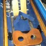
Image from page 778 of “The diseases of infancy and childhood” (1910)
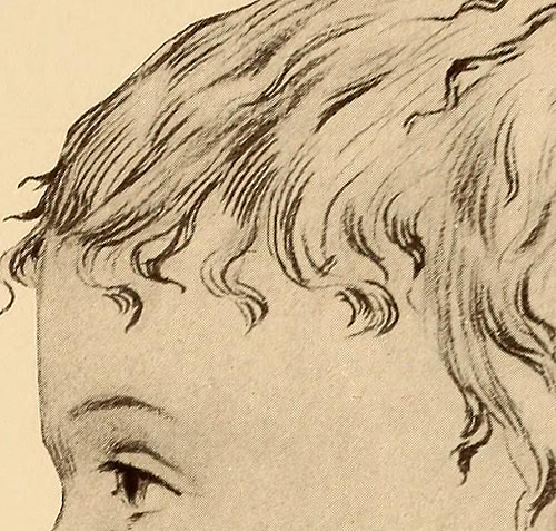
Image by Internet Archive Book Images
Identifier: diseasesofinfa00kopl
Title: The diseases of infancy and childhood
Year: 1910 (1910s)
Authors: Koplik, Henry, 1858- [from old catalog]
Subjects: Children
Publisher: New York and Philadelphia, Lea & Febiger
Contributing Library: The Library of Congress
Digitizing Sponsor: The Library of Congress
View Book Page: Book Viewer
About This Book: Catalog Entry
View All Images: All Images From Book
Click here to view book online to see this illustration in context in a browseable online version of this book.
Text Appearing Before Image:
d, no albumin, no sugar. Microscopicalexamination negative. A series of quantitative urea tests were made in this case. Thegeneral consensus of opinion is that in cases of diabetes insipidus theamount of solids, including the urea, is increased. The tests weremade with the Doremus ureometer. A control test was always made.The table shows marked diminution in the amount of urea. In orderto avoid error, fresh bromine was used. Treatment.—The treatment has been successful in some respects.The child was at once put on a general diet. Antipyrin was given.After the first few days there seemed to be an abatement of the ner-vous symptoms and slight diminution in polydipsia, but no permanentimprovement. He was then given opium several weeks without result.Ergot was next given, and continued for about two months; under thistreatment the pain on the right side disappeared; the restlessnessbecame less, and the thirst likewise diminished. Under a generousdiet the child held his own. PLATE XXX 12
Text Appearing After Image:
1 4 5 I l i. fMm mm / -i* 10 Topography of Enlarged Lymph Nodes. 1. Preauricular nodes enlarged, with disease of the externa] auditory canal, or any eruption on the face, of parotitis 2. Tonsillar nodes. 3. Submaxillary nodes enlarged, with disease of the mouth, or skin eruptions over the lower jaw . 4. Submental nodes enlarged, with chin eruptions, 5. Retropharyngeal nodes enlarged, with infections o the pharynx and the retropharynx. 6. Nodes behind the border of the trapezius muscle enlarged, with disease of the scalp. 7. Nodes behind posterior border of the sternomastoid muscle enlarged, with infections of the retropharynx or the scalp.S. Postaurioular nodes enlarged, with mastoid disease or scalp infections. 9. Nodes above and behind the clavicle enlarged, with infections of the neck or mediastinum.H). Nodes enlarged in infections ,,f the hand or in eruptions such as those o( syphilis. 11. Axillary nodes enlarged, with infections of the arm. the axilla, and the upper chest. 12.
Note About Images
Please note that these images are extracted from scanned page images that may have been digitally enhanced for readability – coloration and appearance of these illustrations may not perfectly resemble the original work.