Some cool Weight loss images:
Image from page 320 of “A treatise on orthopedic surgery” (1910)

Image by Internet Archive Book Images
Identifier: treatiseonorthop1910whit
Title: A treatise on orthopedic surgery
Year: 1910 (1910s)
Authors: Whitman, Royal, 1857-
Subjects: Orthopedics
Publisher: Philadelphia and New York, Lea & Febiger
Contributing Library: Columbia University Libraries
Digitizing Sponsor: Open Knowledge Commons
View Book Page: Book Viewer
About This Book: Catalog Entry
View All Images: All Images From Book
Click here to view book online to see this illustration in context in a browseable online version of this book.
Text Appearing Before Image:
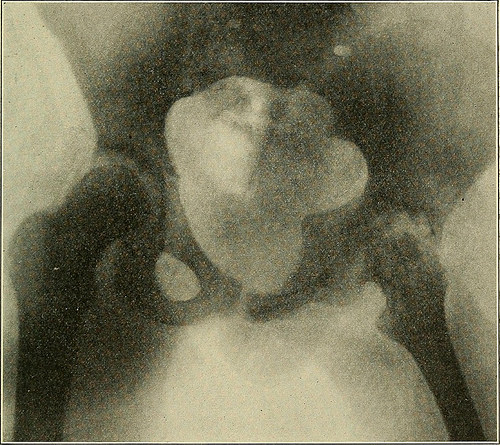
Advanced disease, showing wandering of tbe acetabulum and the obliquity ofthe pelvis due to adduction. Actual shortening one inch, apparent shorteningthree inches. the degree of final inequality depending upon the severity ofthe disease, the duration of the treatment, and upon the impair-ment of function. But even when free motion in the joint isretained, a certain degree of atrophy always persists and the lossin growth is never regained. If motion is completely lost the TUBERCULOUS DISEASE OF THE HIP-JOINT. 323 muscles about the joint lose in bulk in proportion to the disuseof their normal function; whereas the bones of the limb whichare still used to support the weight retain to a greater degreetheir normal size and length. Contrasted with this atrophythere is a relative hypertrophy of the sound limb, which isforced to assume more than its share of work. Actual Shoktenijstg.—^Actual shortening of the limb is aneffect rather than a diagnostic symptom of hip disease. Fig. 223.
Text Appearing After Image:
Illustrating the destructive type of hip disease. The limb having been fixed inabduction. No displacement is present. The causes of actual shortening may be classified as: 1. Disuse of the limb. 2. The effect of the disease upon the epiphyseal cartilage ofthe head of the femur. 3. The more general destructive effects of the disease thatcause upward displacement of the femur. (a) Erosion of the head. (&) Erosion of the acetabulum. 324 OBTHOPEDIC SUBGEEY. (c) Depression of the neck of the femur. (d) Dislocation. Disuse, throughout a long i^eriocl of treatment, causes a cer-tain amount of shortening of the entire limb. To this theshortening of the bones of the leg and of the foot may be attrib-uted in great part. If the epiphyseal cartilage uniting theneck and the head of the femur is destroyed in whole or in partor if the disease hastens union at this point, a certain loss ofgrowth must follow. This is, of course, slight in degree, becausegrowth here is relatively unimportant compar
Note About Images
Please note that these images are extracted from scanned page images that may have been digitally enhanced for readability – coloration and appearance of these illustrations may not perfectly resemble the original work.
Image from page 332 of “Journal of radiology” (1920)
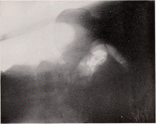
Image by Internet Archive Book Images
Identifier: journalofradiolo1192radi
Title: Journal of radiology
Year: 1920 (1920s)
Authors: Radiological Society of North America American College of Radiology and Physiotherapy
Subjects:
Publisher: Omaha, Neb., : [Radiological Pub. Co.]
Contributing Library: The College of Physicians of Philadelphia Historical Medical Library
Digitizing Sponsor: The College of Physicians of Philadelphia and the National Endowment for the Humanities
View Book Page: Book Viewer
About This Book: Catalog Entry
View All Images: All Images From Book
Click here to view book online to see this illustration in context in a browseable online version of this book.
Text Appearing Before Image:
Case 21. No. 3630The illustration is lateral view. Patient lying face upward. The history shows evidence of digestive disturbance for the past seven years—exaggeratedfor the past three months. Distress immediately upon eating. Loss of weight and strength. Physical examination elicits areas of extreme tenderness in the epigastrium. No tumormass. Barium meal shows constant filling defect in the stomach near the pylorus and theabsence of a normal duodenal cap. The peritoneoscope shows the presence of small metastasis in the peritoneum over the sur-face of the liver and in the gastro-colic omentum. The falciform ligament appears as a denseinfiltrated cord larger than a finger, for some distance away from the anterior abdominal wall,at which point it becomes a thin membrane, whose peritoneum shows the usual glisteningappearance. Diagnosis: Malignant metastasis from gastric carcinoma involving the stomach, duodenum,liver, omentum, etc. PEITONEOSCOPE — ORNDOFF 325
Text Appearing After Image:
Case 34. No. 4040Illustration is lateral view—patient lying left side upward—hips elevated.The history indicates menstruation absent for the past three periods. The oxygen fills the true pelvis and the pelvic brim may be clearly outlined. The uterinedensity is nicely identified by the presence of the round ligament. The size of the uterus ascompared with the pelvic corresponds favorably to approximately the fourth month of preg-nancy. The contour is regular and the shape and position are normal. Contrast the shape with Case 41 which is not normal. Diagnosis: Normal pregnancy approaching the fourth month. ORAL INFECTION IN ITS RELATION TO SYSTEMIC DISEASES* Bex L. Diveley, M. D.and W. W. Duke, M. D. Our work on this subject was started in 1915, and sincethat time we have taken routine dental films on every pa-tient who came into our hands. The series comprised some1800 cases. Each patient received a thorough physical,laboratory and roentgen examination, so we feel certainthat every
Note About Images
Please note that these images are extracted from scanned page images that may have been digitally enhanced for readability – coloration and appearance of these illustrations may not perfectly resemble the original work.
Community Weight Loss Challenge

Image by johnmuk
Photo courtesy of Joanna Stewart, Communications Team, Walsall Council – photo taken at the Family Fit ‘n’ Fun Day on Saturday, 11th May, 2013

Related Posts
 Image from page 666 of “A treatise on orthopedic surgery” (1903)
 Image from page 679 of “A practical treatise on medical diagnosis for students and physicians” (1904)
 Image from page 50 of “A manual of diseases of the nervous system” (1907)
 Image from page 207 of “The Street railway journal” (1884)
 Image from page 150 of “Animals of the past, an account of some of the creatures of the ancient world” (1913)
 Image from page 1263 of “The American florist : a weekly journal for the trade” (1885)